Snoring: is this noisy behavior just an embarrassing situation or is it a sign of an underlying problem? Why obstructive sleep apnoea will almost always leads to loud and frequent snoring, while snoring does not always indicate sleep apnoea? If you suffer of any of these two conditions, it is better that you understand where snoring and sleep apnoea originates because your health is at risk!

While obstructive sleep apnoea is taken more seriously, snoring is usually considered as a simple harmless embarrassing condition. The reality is that both have the same underlying cause that should not be ignored: a poor craniofacial development.

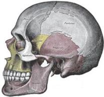
Posture and activity of oral and facial muscles (tongue, masseter, buccinator and others), together with forces coming from the teeth,  determine the direction of growth of the maxilla (Figure 1) [1,2]. Open-mouth posture is one of the most common disturbances of oral posture, always present in mouth breather. However, open-mouth habits do not necessarily coincide with mouth breathing [3].

FacialGrowth
Figure 1 – Direction of growth of the maxilla depends from the forces coming from teeth, tongue and other orofacial muscles. (From [34])
As also confirmed by experiments on primates [4], when the tongue posture is low and teeth are not in contact (at rest and when swallowing), the maxilla drops down and back. This reduces the eye support, flattens the cheekbones, narrows the nasal airway, lengthens the mid facial third, and lowers the palate, which narrows and create malocclusion [5]. Also, a vertical growth of the maxilla forces the mandible to swing back, restricting the pharynx (Figure 2). As a compensatory mechanism, a retruded mandible causes the head to tilt forward in a forward head posture, freeing in this way the airways in the erect posture [6,7].

airwaysOSA
Figure 2 – Two patients from study [8]: subject HB003 shows vertical growth of maxilla, with facial retrognathism  and large mandibular inclination. Notice the forward head posture and the reduced airways’ space. Subject HB092 shows forward growth of maxilla, with facial prognathism and small mandibular inclination. Notice the head posture aligned with the cervical column and the bigger space for the airways.
Restriction of the upper respiratory tract, including narrowing of both nasal cavity and pharinxis a key element in the development of snoring and obstructive sleep apnoea [9,10,11]. In adults, the site of airway occlusion during apnoeic episodes is usually located in the oropharyngeal region. As Figure 3 shows, individuals with a vertical growth of the maxilla have a lower palate, the tongue is in a low posture nearer to the airways and the posterior pharyngeal wall is restricted [12,13,14]. When sleeping lied down, the tongue, the soft palate and the mandible fall into the airways, obstructing the air flow.
tongue-posture
Figure 3 – Individuals with a low tongue posture have a vertical growth of the maxilla, with a lower palate and a retruded mandible. When sleeping lied down, the tongue, the soft palate and the mandible fall into the airways, obstructing the air flow. (From [35])

When airways obstruction is only partial, the air can still flow, but it encounters more resistance, inducing the typical noise of snoring. Instead, when the obstruction is total, the air cannot flows and apnoeic episodes arises, as Figure 4 suggests. As we can seen, the principle is the same for both snoring and apnoea, as well as the underlying cause (a vertical growth of the maxilla).

BlockedAirways
Figure 4 – Partial obstruction of the airways leads to snoring, while total obstruction causes sleep apnoea. (From [36])
Obstructive apnoeas may induce severe intermittent hypoxemia (low level of oxygen in the blood) and carbon dioxide (CO2) retention during sleep, disrupting the normal physiologic interactions between sleep and the cardiovascular system. Hypoxia (deficiency in the amount of oxygen reaching the tissues) and hypercapnia (abnormally elevated carbon dioxide levels in the blood) stimulate ventilatory effort and ultimately arousal from sleep to terminate the apnoeic event. Toward the end of apnoeic episodes, blood pressure can reach levels as high as 240/130 mm Hg (normal blood pressure while awake is between 120/80 and 140/90). For these reasons obstructive sleep apnea is considered a risk factor and possible cause of cardiovascular diseases [15,16,17], but the mechanisms behind the link are unclear.

Hypoxemia caused by sleep apnoea and poor quality of sleep are also linked with attention and memory deficits, resulting as a risk factor for dementia and Alzhaimer’s disease later in life [18,19]. However, also in this case, mechanisms behind this link are not clear.

Snoring and sleep apnoea are also associated with higher incidence of stroke and death [20,21,22], together with hypertension (high blood pressure) [23,24]. Hypertension in mid-life is linked to development of dementia and Alzhaimer’s disease later in life [25,26,27,28]. Link’s origins are unknown in this case too.

So, it looks like there is a clear link between snoring, sleep apnoea, hypertension, cardiovascular diseases, dementia, Alzheimer, stroke and death. Current medicine cannot explain the reasons behind these connections, but the most careful reader should have some doubts.

A poor craniofacial development leads to body’s compensations (e.g. forward head posture, kyphosis, lordosis, scoliosis).  Muscle chains of the body are in continue tension since out of balance, the spine has excessive curvatures and vertebrae are out of alignment. All of this can cause compression of blood vessels, with consequent hypoxemia, hypoxia, as in the case of hair loss, and hypertension. Interestingly, many studies have associated baldness with higher risk of coronary heart disease, of which hypertension is a risk factor [29,30,31,32,33]. So, knowing the common underlying cause of both baldness and obstructive sleep apnea, could craniofacial development be the responsible of all these unclear links?


References

[1] Kiliaridis, Stavros, Christina Mejersjö, and Birgit Thilander. “Muscle function and craniofacial morphology: a clinical study in patients with myotonic dystrophy.” The European Journal of Orthodontics 11.2 (1989): 131-138.

[2] Mew, John RC. “The postural basis of malocclusion: a philosophical overview.” American journal of orthodontics and dentofacial orthopedics 126.6 (2004): 729-738.

[3] Vig, Peter S., et al. “Quantitative evaluation of nasal airflow in relation to facial morphology.” American journal of orthodontics79.3 (1981): 263-272.

[4] Harvold, Egil P., George Chierici, and Karin Vargervik. “Experiments on the development of dental malocclusions.” American journal of orthodontics 61.1 (1972): 38-44.

[5] Mew, M. “Craniofacial dystrophy. A possible syndrome?.” British dental journal 216.10 (2014): 555-558.

[6] Vig, Peter S., Kevin J. Showfety, and Ceib Phillips. “Experimental manipulation of head posture.” American Journal of Orthodontics 77.3 (1980): 258-268.

[7] Gonzalez, Humberto E., and Arturo Manns. “Forward head posture: its structural and functional influence on the stomatognathic system, a conceptual study.” CRANIO® 14.1 (1996): 71-80.

[8] Solow, Beni, and Antje Tallgren. “Head posture and craniofacial morphology.” American Journal of Physical Anthropology 44.3 (1976): 417-435.

[9] CISTULLI, Peter A. “Craniofacial abnormalities in obstructive sleep apnoea: implications for treatment.” Respirology 1.3 (1996): 167-174.

[10] Solow, Beni, et al. “Airway dimensions and head posture in obstructive sleep apnoea.” The European Journal of Orthodontics 18.1 (1996): 571-579.

[11] Nishimura, Tadao, and Kenji Suzuki. “Anatomy of oral respiration: morphology of the oral cavity and pharynx.” Acta oto-laryngologica. Supplementum 550 (2003): 25-28.

[12] Andersson, Lars, and Viveca Brattström. “Cephalometric analysis of permanently snoring patients with and without obstructive sleep apnea syndrome.” International journal of oral and maxillofacial surgery 20.3 (1991): 159-162.

[13] CISTULLI, Peter A. “Craniofacial abnormalities in obstructive sleep apnoea: implications for treatment.” Respirology 1.3 (1996): 167-174.

[14] Nishimura, Tadao, and Kenji Suzuki. “Anatomy of oral respiration: morphology of the oral cavity and pharynx.” Acta oto-laryngologica. Supplementum 550 (2003): 25-28.

[15] Shamsuzzaman, Abu SM, Bernard J. Gersh, and Virend K. Somers. “Obstructive sleep apnea: implications for cardiac and vascular disease.” Jama 290.14 (2003): 1906-1914.

[16] Parish, James M., and Virend K. Somers. “Obstructive sleep apnea and cardiovascular disease.” Mayo Clinic Proceedings. Vol. 79. No. 8. Elsevier, 2004.

[17] Somers, Virend K., et al. “Sleep apnea and cardiovascular disease: An american heart association/american college of cardiology foundation scientific statement from the american heart association council for high blood pressure research professional education committee, council on clinical cardiology, stroke council, and council on cardiovascular nursing in collaboration with the national heart, lung, and blood institute national center on sleep disorders research (national institutes of health).” Circulation 118.10 (2008): 1080-1111.

[18] Johnson, Dayna A., et al. “Greater Cognitive Deficits with Sleep-disordered Breathing among Individuals with Genetic Susceptibility to Alzheimer Disease. The Multi-Ethnic Study of Atherosclerosis.” Annals of the American Thoracic Society14.11 (2017): 1697-1705.

[19] Aoki, Kiyoaki, et al. “Association of sleep‐disordered breathing with decreased cognitive function among patients with dementia.” Journal of sleep research 23.5 (2014): 517-523.

[20] Palomäki, Heikki, et al. “Snoring, sleep apnea syndrome, and stroke.” Neurology 42.7 Suppl 6 (1992): 75-81.

[21] Dyken, Mark E., et al. “Investigating the relationship between stroke and obstructive sleep apnea.” Stroke 27.3 (1996): 401-407.

[22] Yaggi, H. Klar, et al. “Obstructive sleep apnea as a risk factor for stroke and death.” New England Journal of Medicine353.19 (2005): 2034-2041.

[23] Nieto, F. Javier, et al. “Association of sleep-disordered breathing, sleep apnea, and hypertension in a large community-based study.” Jama 283.14 (2000): 1829-1836.

[24] Marcus, Carole L., Mary G. Greene, and John L. Carroll. “Blood pressure in children with obstructive sleep apnea.” American journal of respiratory and critical care medicine157.4 (1998): 1098-1103.

[25] McGrath, Emer R., et al. “Blood pressure from mid‐to late life and risk of incident dementia.” Neurology (2017): 10-1212.

[26] Skoog, Ingmar, et al. “15-year longitudinal study of blood pressure and dementia.” The Lancet 347.9009 (1996): 1141-1145.

[27] Skoog, Ingmar, and Deborah Gustafson. “Update on hypertension and Alzheimer’s disease.” Neurological research28.6 (2006): 605-611.

[28] Kivipelto, Miia, et al. “Midlife vascular risk factors and Alzheimer’s disease in later life: longitudinal, population based study.” Bmj 322.7300 (2001): 1447-1451.

[29] Yamada, Tomohide, et al. “Male pattern baldness and its association with coronary heart disease: a meta-analysis.” BMJ open 3.4 (2013): e002537.

[30] Sharma, Kamal H., and Anchal Jindal. “Association between androgenetic alopecia and coronary artery disease in young male patients.” International journal of trichology 6.1 (2014): 5.

[31] Sharma, Lata, et al. “Androgenetic alopecia and risk of coronary artery disease.” Indian dermatology online journal4.4 (2013): 283.

[32] Lotufo, Paulo A., et al. “Male pattern baldness and coronary heart disease: the Physicians’ Health Study.” Archives of internal medicine 160.2 (2000): 165-171.

[33] Herrera, Carlos R., et al. “Baldness and coronary heart disease rates in men from the Framingham Study.” American journal of epidemiology 142.8 (1995): 828-833.

Other websites

[34] John Mew’s Legacy, Mystery of Crooked Teeth Unraveled, by Claiming Power

[35] Maxilla, single most important bone in the body?, by Claiming Power

[36] Snoring, Sleep Apnea and Dentistry, by Design for Dental Health Blog